Ken Unruh MD hand surgeon with patient

Navigating Hand & Wrist Pain: A Guide to Treatment Options

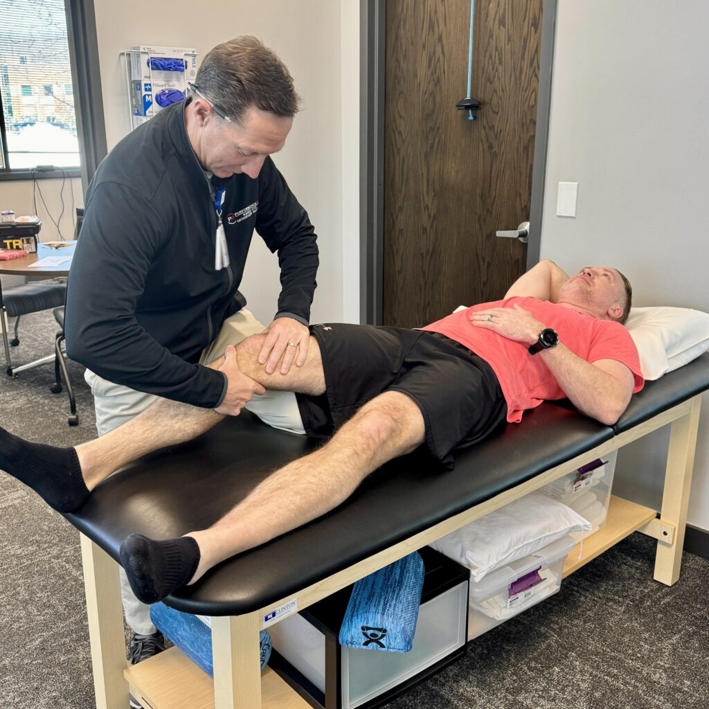
That persistent ache in your hand, the tingling sensation in your fingers, the stiffness that makes it difficult to grip a coffee mug, or the lingering pain from a sprained wrist can be more than just an inconvenience. Hand pain and wrist pain can significantly impact your daily life, making simple tasks feel frustrating and difficult. We understand that finding effective relief is key to restoring a pain-free and active lifestyle.

At Kansas City Orthopaedic Institute (KCOI), our dedicated team of orthopedics experts understands the intricate anatomy of the hand and wrist. We specialize in diagnosing and treating a wide range of conditions, from common overuse issues, like tendonitis, to complex injuries, such as fractures or dislocations. This blog post will serve as your comprehensive guide to understanding both the surgical and non-surgical treatment options available for hand pain and wrist pain, helping you navigate your path to recovery.

Understanding the Sources of Hand and Wrist Pain

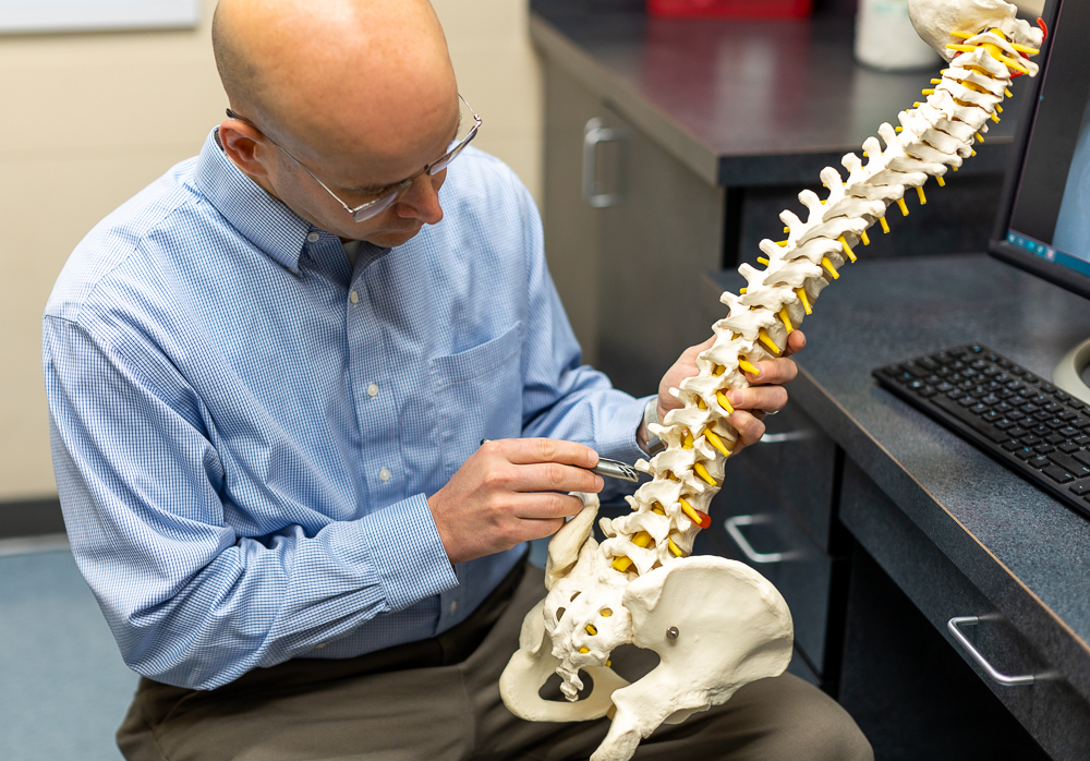
The hand and wrist are marvels of anatomical engineering, composed of 27 bones, a complex network of ligaments, tendons, muscles, and nerves. Due to their frequent use and delicate structure, they are susceptible to a variety of conditions and injuries. The most common causes of hand pain and wrist pain include:

  • Carpal Tunnel Syndrome: This is a condition where the median nerve, which runs from the forearm into the hand, becomes compressed at the wrist. It’s often caused by repetitive motions, forceful gripping, and exposure to high vibration activities.  It can lead to tingling, numbness, weakness, and pain in the hand and fingers.
  • Osteoarthritis: Often called “wear-and-tear” arthritis, it occurs when the cartilage that cushions the joints breaks down, leading to pain, stiffness, and loss of motion. It commonly affects the base of the thumb and the small joints of the fingers.
  • Tendonitis and Tenosynovitis: Inflammation of a tendon or its sheath, often resulting from repetitive stress or overuse. A prime example is trigger finger, where a finger or thumb gets stuck in a bent position.
  • Fractures: A break in one of the small bones of the hand or wrist, such as a wrist fracture from a fall.
  • Sprained Wrist: A common injury that occurs when the ligaments supporting the wrist are stretched or torn, often from a fall, sports activity, or sudden impact. It can cause swelling, bruising, and pain with movement.
  • Ganglion Cysts: Noncancerous, fluid-filled lumps that most often develop on the back of the wrist.
  • De Quervain’s Tenosynovitis: Inflammation of the tendons on the thumb side of the wrist, causing pain with gripping or pinching motions.
orthopedic surgeon examining a patient's arm with tool


Non-Surgical Treatment Options: The First Step to Relief

For many patients, especially those with mild to moderate symptoms, the journey to relief begins with conservative, non-surgical treatments. These approaches focus on reducing pain and inflammation, restoring function, and preventing further irritation. Our orthopedics team prioritizes these options whenever possible.

  • Rest and Activity Modification: The simplest and often most effective treatment for overuse injuries. Avoiding or modifying activities that aggravate your symptoms can give the tissues time to heal.
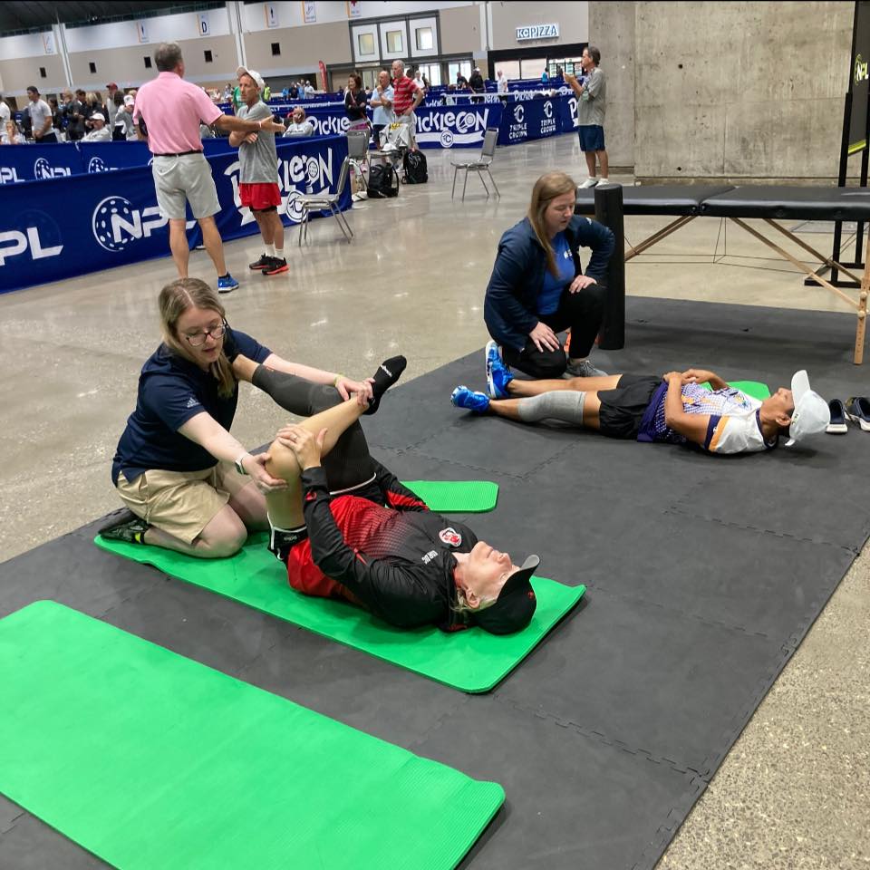
  • Physical or Occupational Therapy: A certified therapist can provide a personalized plan of exercises to improve strength, flexibility, and range of motion. They may also teach you ergonomic techniques to reduce strain during daily activities.
  • Bracing or Splinting: Wearing a brace or splint can stabilize the hand or wrist, reducing stress on tendons and nerves and allowing inflamed tissues to rest and heal. This is particularly helpful for carpal tunnel syndrome, wrist tendonitis, or a sprained wrist.
  • Medications: Over-the-counter anti-inflammatory medications, such as ibuprofen, can help manage pain and reduce swelling.
  • Therapeutic Injections: A steroid injection can be a powerful tool to provide significant, though often temporary, relief from pain and inflammation. For conditions like carpal tunnel syndrome, therapeutic injections can reduce swelling around the compressed nerve. They are also effective for relieving the pain of arthritis and trigger finger.

When to Consider Surgery: A Targeted Approach

When non-surgical treatments no longer provide adequate relief, or in cases of severe conditions like a complex wrist fracture, surgery becomes the most effective option. Modern orthopedic surgery for the hand and wrist is highly specialized and often minimally invasive, with a focus on restoring long-term function and alleviating pain.

The expert surgeons at KCOA, our partner physician clinic, utilize the latest techniques to ensure the best possible outcomes for our patients. The following physicians constitute the hand and wrist team at KCOI, and specialize in surgical and non-surgical treatment for hands and wrists:

Dr. Mark Winston examining a patient's hand

Here are some of the most common hand and wrist surgical procedures:

Carpal Tunnel Release

This is one of the most common hand surgeries. The goal is to relieve the pressure on the median nerve. The procedure involves cutting the transverse carpal ligament, which forms the “roof” of the carpal tunnel, to create more space for the nerve. The ligament eventually heals in a way that allows more room, preventing future compression. This procedure has a very high success rate in relieving symptoms of tingling, numbness, and weakness. It can be performed through a traditional open incision or an endoscopic, minimally invasive technique, often with a faster recovery time.

Trigger Finger A1 Pulley Release

Trigger finger occurs when the A1 pulley, a small fibrous sheath that holds the tendon close to the bone, becomes inflamed and thickens, making it difficult for the tendon to glide smoothly. This causes the characteristic “catching” or “locking” sensation. The surgical procedure, trigger finger A1 pulley release, is a simple but highly effective outpatient procedure. The surgeon makes a small incision and carefully cuts the A1 pulley, releasing the tendon. The pulley eventually heals in an open position, allowing the tendon to glide freely and restoring normal finger movement.

Wrist Arthroscopy

Arthroscopy is a minimally invasive surgical technique that uses a small camera (arthroscope) to view and operate inside a joint. For the wrist, this allows the surgeon to diagnose and treat a variety of conditions without a large incision. Wrist arthroscopy can be used to:

  • Repair torn ligaments and cartilage (e.g., TFCC tears).
  • Remove inflamed tissue.
  • Remove or repair bone fragments.
  • Debride or clean up the joint from arthritis.

This technique leads to less pain, minimal scarring, and a faster recovery compared to traditional open surgery.

Fracture Repair

A wrist fracture can be a painful and debilitating injury. While some simple fractures can be treated with a cast or splint, more complex fractures—especially those that are displaced or involve the joint—require surgery. The goal of fracture repair is to realign the broken bones and stabilize them with plates, screws, or pins. This allows the bone to heal in the correct position, restoring proper wrist alignment and function. A dedicated orthopedics specialist is essential for managing these types of injuries.

physician reviewing hand xray with a patient

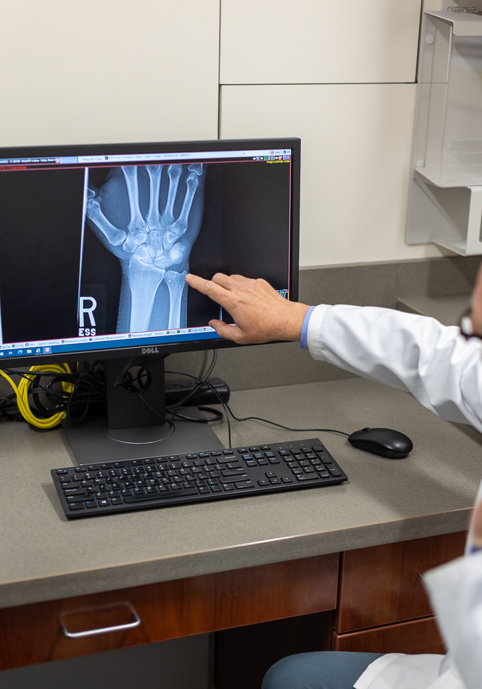
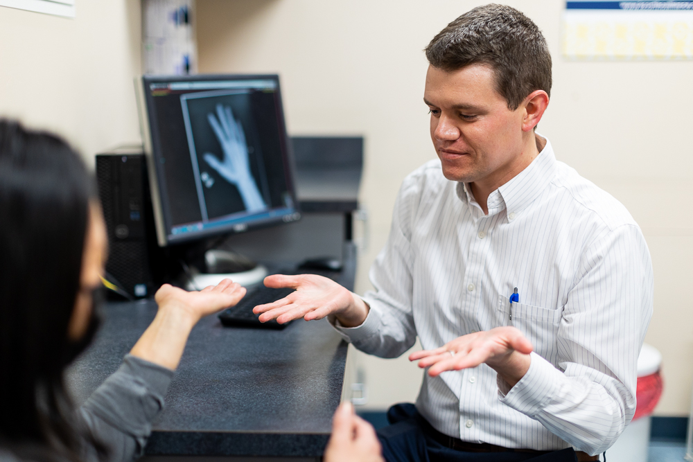
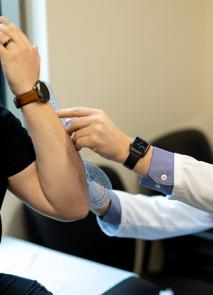
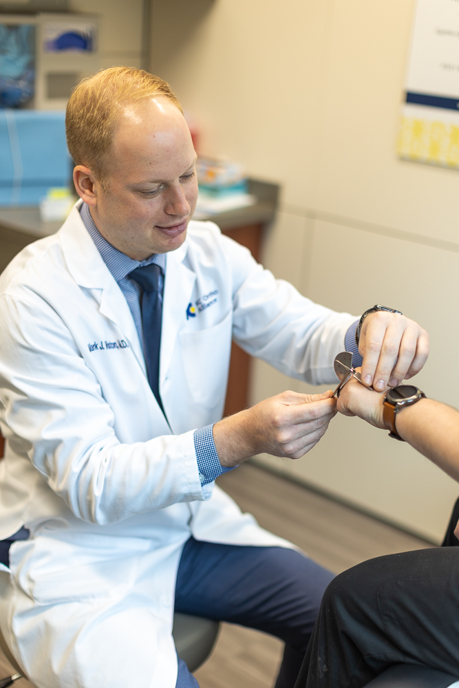
The Importance of a Comprehensive Diagnosis

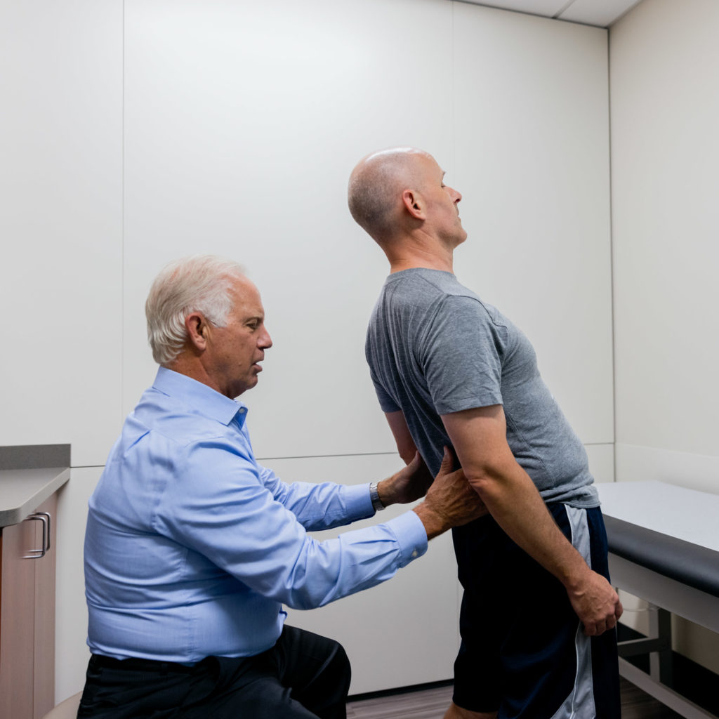
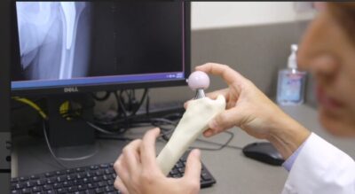
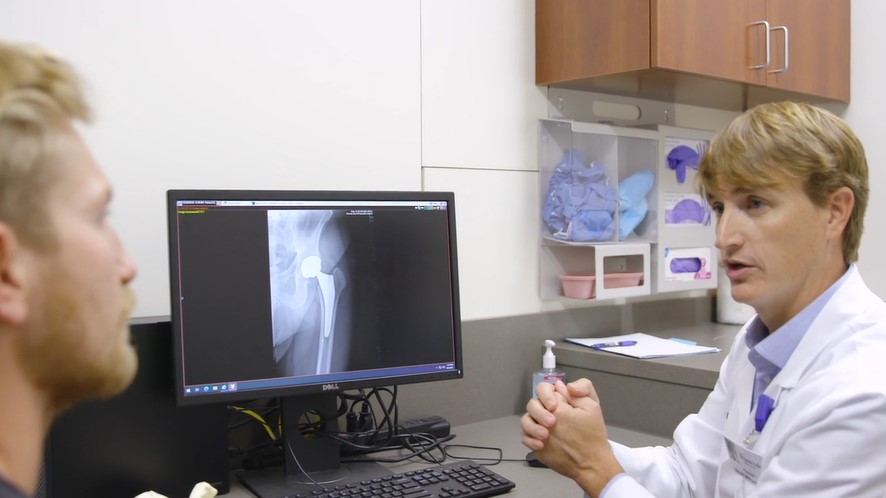
Regardless of your symptoms, the most crucial first step is a proper diagnosis. Our specialists perform a thorough evaluation that includes a physical exam, a review of your medical history, and may include diagnostic imaging such as X-rays, MRI, or ultrasound. This detailed assessment allows our team to pinpoint the exact source of your hand pain or wrist pain and develop a personalized treatment plan that is right for you.

You can learn more about our specialized upper extremity services and hand and wrist care on our website. Our team includes fellowship-trained surgeons with extensive experience in the latest surgical and non-surgical techniques, ensuring you receive the highest level of care.

Take the First Step Towards a Pain-Free Future

Don’t let hand pain and wrist pain hold you back from doing the things you love. Whether your symptoms are mild or severe, there are effective solutions available. From simple therapeutic injections to advanced surgical procedures like carpal tunnel release and wrist arthroscopy, our hand and wrist team at KCOI is dedicated to helping you find relief and restore your quality of life.

We are proud to serve the Kansas City metropolitan area and surrounding communities with compassionate and expert orthopedic care. Take control of your health and schedule a consultation to discuss your treatment options.

Ready to find relief? Contact the expert orthopedics team at Kansas City Orthopedic Alliance today: Request an Appointment. Your path to recovery starts here.

Back Pain & Stiffness: Find Relief, Restore Your Life

Back pain affects people of all ages and can disrupt daily life, from work to the activities you enjoy. At Kansas City Orthopaedic Institute (KCOI), our specialists are here to help you understand the cause of your pain and explore personalized treatment options. In this blog, we’ll break down the most common causes and solutions to help you find lasting relief.

Understanding Your Back: A Marvel of Engineering

Understanding back pain starts with the spine—a complex structure of 33 vertebrae, cushioned by discs, connected by ligaments, and supported by muscles. It protects the spinal cord and enables movement. When any part is irritated or damaged, especially in the lower back, it can lead to pain, stiffness, and other symptoms.

The Many Faces of Back Pain: Common Causes

Back pain can stem from a variety of sources, ranging from simple muscle strains to more complex degenerative conditions. Recognizing the potential causes is the first step toward effective treatment.

  • Muscle Strains and Ligament Sprains: Often caused by sudden awkward movements, lifting heavy objects improperly, or overstretching during physical activity. These are common injuries, especially in sports medicine.
  • Disc Problems:
    • Herniated or Bulging Discs: Over time, the discs can degenerate, and their soft inner material can push out (bulge) or rupture through the outer layer (herniate), pressing on nearby nerves. This is a very common cause of lower back pain and can lead to sciatica (pain radiating down the leg).
    • Degenerative Disc Disease: As we age, discs naturally lose hydration and elasticity, becoming less effective shock absorbers and leading to pain and stiffness.
  • Arthritis (Osteoarthritis): The cartilage cushioning the facet joints (small joints connecting the vertebrae) can wear down, leading to bone-on-bone friction, inflammation, and pain. This is a common cause of chronic back pain.
  • Spinal Stenosis: A narrowing of the spinal canal, which can put pressure on the spinal cord and nerves. This often causes pain, numbness, or weakness in the legs, especially when walking.
  • Spondylolisthesis: Spondylolisthesis, a condition where a vertebra slips forward, is a concern for young athletes, often stemming from spondylolysis, a stress fracture in the spine. This condition is frequently caused by repetitive twisting and hyperextension movements, making it prevalent in sports like gymnastics and football.
  • Fractures: Trauma (like a fall or car accident) or weakened bones (due to osteoporosis) can lead to vertebral fractures.
  • Scoliosis: An abnormal curvature of the spine, which can cause pain and imbalance, especially if severe.
  • Lifestyle Factors: Poor posture, prolonged sitting, lack of exercise, obesity, and smoking can all contribute to developing or worsening back pain.
Dr. Ted Koreckij and spine model

When to See an Orthopedic Specialist for Your Back Pain

Many instances of back pain are acute, meaning they come on suddenly and resolve within a few days or weeks with rest and self-care. However, certain symptoms warrant immediate attention from an orthopedic specialist. Don’t delay seeking professional help if you experience:

  • Severe pain that doesn’t improve with rest.
  • Pain radiating down one or both legs, especially if it extends below the knee.
  • Numbness, tingling, or weakness in your legs, feet, or groin area.
  • Loss of bowel or bladder control (a medical emergency).
  • Unexplained weight loss accompanied by back pain.
  • Fever with back pain.
  • Pain that is worse at night or awakens you from sleep.
  • Pain following a significant injury, such as a fall or car accident.

Even if your symptoms aren’t severe or alarming, if your back pain persists for more than a few weeks, interferes with your daily activities, or you find yourself constantly battling stiffness, it’s time to consult with an expert. Our team at KCOI is equipped to provide a comprehensive evaluation, diagnose the precise cause of your pain, and recommend the most effective treatment plan tailored to your needs.

Navigating Non-Surgical Treatment Options: The First Line of Defense

For most patients, the journey to relief from back pain begins with non-surgical, conservative treatments. These approaches focus on reducing pain, improving function, and empowering you to manage your condition effectively. At KCOI, we believe in a multidisciplinary approach, often combining several therapies for optimal results.

  • Rest and Activity Modification: In the acute phase, brief rest can be beneficial. However, prolonged bed rest is generally discouraged. Your doctor may recommend modifying certain activities to avoid movements that aggravate your pain.
  • Physical Therapy: This is often the cornerstone of non-surgical back pain treatment. A skilled physical therapist can teach you specific exercises to strengthen core muscles, improve flexibility, correct posture, and enhance body mechanics. They can also use modalities like heat, ice, ultrasound, or electrical stimulation to reduce pain and inflammation. Through our partership with KCOI Performance Rehab, there are 10 convenient outpatient locations for PT and OT.
  • Medications:
    • Over-the-counter pain relievers: NSAIDs (like ibuprofen or naproxen) can reduce pain and inflammation.
    • Muscle relaxants: May be prescribed for muscle spasms.
    • Neuropathic pain medications: For nerve-related pain.
  • Injections:
    • Epidural Steroid Injections: These target inflammation around irritated spinal nerves, providing significant, though often temporary, pain relief.
    • Facet Joint Injections: Address pain stemming from the small facet joints in the spine.
    • Nerve Blocks: Can identify the source of pain or provide sustained relief by blocking nerve signals.
  • Chiropractic Care and Manual Therapy: For some types of back pain, manipulation and mobilization techniques can help restore joint motion and reduce pain.
  • Alternative Therapies: Acupuncture, massage therapy, and yoga can be complementary treatments for some individuals, helping to manage pain and improve flexibility.
  • Lifestyle Changes: Weight management, quitting smoking, and adopting ergonomic practices at work and home are crucial for long-term back health. Our experts can offer guidance on safe exercise and activity.
Dr. Lowry Jones doing back exam

Considering Surgical Intervention: When Conservative Care Isn’t Enough

While the vast majority of back pain cases improve with conservative care, there are situations where surgery becomes a necessary and effective option. Your orthopedic surgeon at KCOI will only recommend surgery after a thorough evaluation, when non-surgical treatments have been exhausted, and if clear diagnostic evidence (like MRI scans) points to a specific anatomical problem that can be corrected surgically.

Surgical intervention for spine conditions aims to:

  • Decompress Nerves: Relieve pressure on compressed nerves that are causing pain, numbness, or weakness.
  • Stabilize the Spine: Fuse vertebrae together or use hardware to prevent excessive movement, particularly for instability or severe deformities.

Common types of spine surgeries for back pain include:

  • Laminectomy: Often performed for herniated discs or spinal stenosis. A laminectomy involves removing part of the vertebral bone (lamina) to create more space for nerves.
  • Discectomy: A discectomy removes the portion of a herniated disc that is pressing on a nerve. These can often be done with minimally invasive techniques.
  • Spinal Fusion: This procedure involves permanently connecting two or more vertebrae using bone grafts and often metal hardware (screws, rods). It’s used to stabilize the spine, correct deformities, or eliminate painful motion between vertebrae, particularly in cases of severe degenerative disc disease, spondylolisthesis, or spinal instability.
  • Artificial Disc Replacement: In some select cases, particularly for single-level degenerative disc disease in younger patients, the damaged disc can be removed and replaced with an artificial disc in the cervical spine, preserving motion at that spinal level.
  • Vertebroplasty/Kyphoplasty: Procedures used to treat painful vertebral compression fractures, often due to osteoporosis. Bone cement is injected into the fractured vertebra to stabilize it.

The choice of surgical procedure depends entirely on the specific diagnosis, the severity of your condition, your overall health, and the recommendations of your orthopedic specialist. Our orthopedics team at KCOI prides itself on utilizing the latest surgical techniques, including minimally invasive options, to ensure the best possible outcomes for our patients.

Prevention and Proactive Spine Health

While we can’t always prevent back pain, especially from degenerative changes, adopting healthy habits can significantly reduce your risk and severity:

  • Maintain a Healthy Weight: Excess weight, particularly in the abdomen, puts added strain on your lower back pain region.
  • Practice Good Posture: Be mindful of how you sit, stand, and lift. When lifting, use your legs, not your back.
  • Stay Active: Regular exercise strengthens core muscles that support your spine. Include activities that promote flexibility and cardiovascular health. Even gentle movement can help prevent stiffness.
  • Ergonomics: Ensure your workstation is ergonomically sound to support your spine during prolonged sitting.
  • Quit Smoking: Smoking reduces blood flow to the discs, accelerating their degeneration.
  • Listen to Your Body: Pay attention to early warning signs of pain and take appropriate rest or modify activities.

Take Control of Your Back Health

Persistent back pain and stiffness don’t have to be your norm. Whether it’s a recent injury or chronic discomfort, our experts provide accurate diagnoses and personalized treatment to relieve pain, restore function, and improve your quality of life. Don’t let back pain hold you back. Request an appointment with one of our experienced orthopedic specialists at partner physician clinic KCOA today: Request an Appointment. Your spine health is our priority.

Dr TJ Rasmussen and knee model

Total vs. Partial Knee Replacement: What You Should Know

This blog post will dive into both total and partial knee replacements, explaining when each might be recommended, and help you understand how our team at KCOI can guide you through this journey.

Understanding Knee Osteoarthritis: The Root of the Problem

The most common reason people consider knee replacement surgery is severe osteoarthritis. This degenerative joint disease occurs when the protective cartilage that cushions the ends of your bones wears away over time. In the knee, this includes the cartilage on the end of your thigh bone (femur), shin bone (tibia), and kneecap (patella).

As this cartilage deteriorates, the bones begin to rub directly against each other, leading to:

  • Pain: Often described as a deep ache, sharp pain, or grinding sensation, especially with movement or after periods of rest.
  • Stiffness: Making it difficult to bend or straighten the knee, particularly in the mornings.
  • Swelling: Due to inflammation within the joint.
  • Limited Range of Motion: Affecting your ability to walk, climb stairs, or perform daily activities.
  • Clicking or Popping Sounds: Known as crepitus, as the joint surfaces grate against each other.

While osteoarthritis is often age-related, previous knee injuries (such as ligament tears from sports or trauma), genetics, obesity, and overuse can also contribute to its development. When these symptoms significantly impact your quality of life and non-surgical treatments like physical therapy, medication, and injections are no longer effective, your orthopedic specialist may discuss surgical intervention.

Non-Surgical Approaches: When Is Surgery the Next Step?

Before recommending surgery, your orthopedic surgeon will always explore a range of non-surgical treatments. Such approaches include:

  • Physical Therapy: Targeted exercises to strengthen the muscles around the knee, improve flexibility, and enhance stability.
  • Medications: Over-the-counter pain relievers (like NSAIDs), prescription anti-inflammatory drugs, or pain medications.
  • Injections: Corticosteroid injections to reduce inflammation and pain, or viscosupplementation injections (hyaluronic acid) to lubricate the joint.
  • Lifestyle Modifications: Weight management to reduce stress on the knee, and activity modifications to avoid high-impact movements that aggravate symptoms.
  • Assistive Devices: Using a cane or walker to reduce load on the affected knee.
  • Compression Sleeves & Braces: Sleeves can reduce swelling and inflammation, while braces recreate stability and offload the knee joint.

You can learn more about comprehensive orthopedic treatments available at KCOI for various knee conditions, including those managed by our skilled sports medicine specialists for less severe injuries.

The decision to move towards knee replacement surgery is made when these conservative methods fail to provide lasting relief, and the pain severely limits your daily activities and quality of life. This is where understanding your surgical options becomes paramount.

Total Knee Replacement (TKR): A Comprehensive Solution

Total Knee Replacement (TKR) relieves severe knee pain and restores function by replacing damaged joint surfaces with metal and plastic implants. At KCOI, we use advanced techniques—including robotic-assisted surgery—to customize each procedure to the patient’s unique anatomy.

How TKR Works:

  • Femoral Component: A metal cap covers the end of the thigh bone.
  • Tibial Component: A flat metal plate with a polyethylene (plastic) insert is placed on top of the shin bone. The plastic insert acts as a new smooth cartilage surface.
  • Patellar Component (Optional): A plastic button may be placed on the underside of the kneecap.

The goal of TKR is to resurface all three compartments of the knee (medial, lateral, and patellofemoral) to eliminate bone-on-bone friction, reduce pain, and improve the knee’s alignment and movement. TKR is often recommended for patients with widespread arthritis affecting multiple areas of the knee joint.

Partial Knee Replacement (PKR): A More Targeted Approach

Partial Knee Replacement (PKR) is a less invasive option for patients with arthritis limited to one knee compartment, typically the inner side. It preserves healthy bone and cartilage by replacing only the damaged area. Dr. Sean Bonanni notes PKRs work well for isolated wear but may not last as long as total knee replacements.

How PKR Works:
  • Only the damaged cartilage and bone from the affected compartment are removed.
  • Small metal components are resurfaced onto the ends of the femur and tibia in that specific area.
  • A plastic insert is placed between these metal components to create a new smooth gliding surface.
Advantages of PKR (when appropriate):
  • Smaller Incision: Generally, a less extensive incision compared to TKR.
  • Less Bone Removal: Preserves more of your natural bone and ligaments.
  • Potentially Faster Recovery: Many patients experience a quicker return to activities.
  • More Natural Feel: Some patients report that a PKR knee feels “more natural” because more of their own knee anatomy is preserved.
  • Reduced Blood Loss: Typically less blood loss during surgery.
  • Shorter Hospital Stay: Often allows for a shorter recovery period in the hospital.
Considerations for PKR:

PKR is not for everyone. Ideal candidates usually have:

  • Arthritis primarily affecting only one compartment of the knee.
  • Intact knee ligaments (especially the ACL).
  • Reasonable knee alignment.
  • No significant inflammation or other widespread knee conditions.

Your orthopedic surgeon will carefully evaluate your specific condition, including X-rays and potentially MRI scans, to determine if you are a suitable candidate.

Choosing the Right Path: Factors Your Orthopedic Surgeon Considers

The decision between a Total Knee Replacement and a Partial Knee Replacement is complex and highly individualized. It involves a thorough discussion with your orthopedic surgeon at KCOI, taking into account several factors:

  • Arthritis Extent: Widespread = TKR; isolated = PKR possible
  • Ligament Stability: Strong ACL needed for PKR
  • Knee Alignment: Major deformities may require TKR
  • Age & Activity: May influence implant choice and recovery
  • Patient Goals: Pain relief and function drive decision
  • Surgeon Expertise: Surgeon selects best fit for your anatomy
  • Bone Quality: Good bone needed for secure implant placement

What to Expect: Recovery and Rehabilitation

Regardless of whether you undergo a total or partial knee replacement, rehabilitation is a critical component of your recovery. Both procedures aim to alleviate pain and restore function, but the journey involves dedication and effort.

  • Hospital Stay: Typically a few days, though some patients may qualify for outpatient joint replacement.
  • Pain Management: Your care team will provide medication and strategies to manage post-surgical pain.
  • Physical Therapy: Begins almost immediately after surgery, focusing on restoring range of motion, strength, and walking ability. This is crucial for optimizing your outcome.
  • Home Recovery: Continuing exercises, managing swelling, and gradually increasing activity levels as advised by your surgeon and physical therapist.

Our commitment at KCOI extends beyond the operating room. We work closely with our partners at KCOA to provide a seamless and comprehensive patient experience. From pre-operative education to post-operative rehabilitation, our goal is to support you every step of the way towards a successful recovery and a pain-free life.

Life After Knee Replacement

Modern knee replacements are highly successful at relieving pain and improving quality of life. Most patients return to daily activities like walking, swimming, or cycling. High-impact sports are usually discouraged, but your surgeon will guide you based on your recovery and procedure type.

Read about KCOA patient Leo Sebus, and his journey from pain to knee replacement to recovery.  Sebus tells us that he relied on his team’s guidance on pain management and routines, and differentiating between pain, soreness and general discomfort helped him navigate his recovery. 


Take the Next Step Towards a Pain-Free Life

Living with chronic knee pain doesn’t have to be your reality. Understanding the options available, from conservative treatments to advanced surgical procedures like Total and Partial Knee Replacement, is the first step towards reclaiming your mobility and improving your quality of life.

At KCOI, our expert surgeons use the latest techniques to treat all knee conditions with personalized care. Don’t let knee pain hold you back.

If you’re experiencing persistent knee pain and want to explore your treatment options, contact KCOI today. Schedule a consultation through the physician clinic at KCOA: Request an Appointment. Your journey to a healthier, more active life starts here.

pickleball players

Pickleball Injuries: When to Seek Orthopedic Care

Pickleball has exploded in popularity, offering a fun and fast-paced blend of tennis, badminton and ping pong. It’s easy to learn, highly social and offers a fantastic way to stay active. However, as with any sport that involves quick movements, pivots and explosive actions, pickleball comes with its own set of potential risks, particularly for the lower extremities.

At Kansas City Orthopaedic Institute (KCOI), our sports medicine specialists and orthopedic surgeons are increasingly seeing patients with pickleball-related injuries. While the game is generally considered low-impact, the sudden starts, stops and directional changes can place significant stress on your feet, ankle, knee, leg and hip. This post will delve into common lower extremity pickleball injuries, explain why they occur, and, most importantly, help you understand when it’s time to transition from self-care to professional orthopedic treatment.

The Rise of Pickleball and the Twist of Injuries

The explosion in popularity (and pickleball courts throughout the KC metro) means more people are playing, often without a prior background in racket sports or high-intensity activity. This broad appeal, while wonderful for public health, can also contribute to a higher incidence of injuries. Players might underestimate the demands the game places on their bodies, leading to improper warm-ups, pushing too hard too fast, or not recognizing the early signs of strain.

Common lower extremity injuries in pickleball frequently stem from:

  • Sudden directional changes: Quickly moving side-to-side or forward-and-back puts immense pressure on ankles and knees.
  • Repetitive impacts: Constant running and jumping on hard courts can stress joints and bones.
  • Overextension or awkward landings: Reaching for a shot or landing off-balance can lead to sprains or strains.
  • Lack of proper conditioning: Not adequately preparing your muscles and joints for the sport’s demands.

While this post focuses on lower extremity injuries, it’s worth noting that other common pickleball injuries can include issues with the wrist (from trip/falls or swings/strokes), elbow and shoulder (excessive swings/strokes).

Common Lower Extremity Pickleball Injuries

Let’s break down some of the specific lower extremity injuries our orthopedic experts at KCOI frequently diagnose and treat:

Ankle Sprains

Perhaps the most common injury in pickleball, an ankle sprain, occurs when the ligaments that stabilize your ankle are stretched or torn. This often happens during quick lateral movements, lunging for a ball, or landing awkwardly after a jump. You might feel a “pop” or tearing sensation, followed by immediate pain, swelling, bruising and difficulty bearing weight.

Achilles Tendinopathy or Rupture

The Achilles tendon, located at the back of your ankle, connects your calf muscles to your heel bone. The explosive push-off movements required in pickleball can put significant strain on this tendon. Overuse can lead to Achilles tendinopathy (inflammation or degeneration), characterized by pain and stiffness in the back of the heel, especially in the morning or after activity. A sudden, forceful push-off can also lead to a complete Achilles tendon rupture, which is often described as feeling like you’ve been “kicked” in the back of the leg, followed by severe pain and inability to push off your foot.

Knee Ligament Sprains (ACL, MCL, LCL) and Meniscus Tears 

The knee is particularly vulnerable due to the twisting and pivoting motions in pickleball.

  • ACL (Anterior Cruciate Ligament) injuries: Often occur with sudden stops, changes in direction, or awkward landings, leading to a “pop” sensation, instability, swelling and severe pain.
  • MCL (Medial Collateral Ligament) injuries: Result from forces that push the knee inward, common during side-to-side movements or collisions.
  • LCL (Lateral Collateral Ligament) injuries: Less common, caused by forces pushing the knee outward.
  • Meniscus Tears: The menisci are C-shaped pieces of cartilage that cushion your knee joint. Twisting the knee while bearing weight can tear a meniscus, causing pain, swelling, clicking, catching or a feeling of the knee “giving out.”

Strains and Fractures

Hamstring Strains

The hamstrings, located at the back of your thigh, are crucial for running and stopping movements. Sudden sprints or lunges, especially without adequate warm-up, can lead to a hamstring strain, causing sharp pain in the back of the thigh, bruising and tenderness.

Calf Strains (e.g., “Tennis Leg”)

Similar to hamstring strains, calf strains (often called “tennis leg” in other sports, but equally applicable to pickleball) occur when the calf muscles are suddenly stretched or contracted beyond their capacity. This can happen during a powerful push-off or an abrupt stop, resulting in sharp pain in the back of the lower leg, swelling and difficulty walking.

Plantar Fasciitis

This condition involves inflammation of the plantar fascia, a thick band of tissue running along the bottom of your foot from your heel to your toes. The repetitive impact of running and jumping on hard pickleball courts, especially with inadequate footwear or foot support, can lead to heel pain that is often worse in the morning or after periods of rest.

Stress Fractures

While less common acutely, repetitive stress on the bones of the foot or lower leg (tibia, fibula) without adequate recovery time can lead to stress fractures. These are tiny cracks in the bone, causing localized pain that worsens with activity and improves with rest.

physical therapy on leg

How to Know When You Need Orthopedic Treatment

It’s natural to have some aches and pains after playing a new sport or pushing yourself during a game. However, certain signs indicate that your injury is more than just a minor tweak and warrants evaluation by a sports medicine physician or orthopedic surgeon at KCOI.

Seek immediate medical attention if you experience:

  1. Sudden, Severe Pain: Pain that is excruciating and prevents you from continuing to play or bear weight.
  2. Deformity: If the injured area looks visibly crooked, swollen or out of place.
  3. Inability to Bear Weight: If you cannot put any weight on your foot or leg without excruciating pain.
  4. Audible “Pop” or “Snap”: Often indicative of a ligament tear (like ACL or Achilles) or a fracture.
  5. Loss of Function: If you cannot move the affected joint or limb normally.
  6. Numbness or Tingling: This could indicate nerve involvement and requires prompt evaluation.

Consider scheduling an appointment with an orthopedic specialist if you have:

  1. Persistent Pain: Pain that doesn’t improve with rest, ice, compression, and elevation (RICE method) after a few days.
  2. Swelling and Bruising that Worsens: Or does not subside after 24-48 hours.
  3. Limited Range of Motion: If you can’t fully straighten or bend your joint compared to the uninjured side.
  4. Instability: A feeling that your knee or ankle might “give out” or is unstable during movement.
  5. Recurring Pain: If the pain keeps coming back every time you play pickleball, even after resting.
  6. Chronic Aches: Persistent aches that disrupt sleep or daily activities, even if not acutely severe.

At KCOI, our team of orthopedic surgeons has extensive experience diagnosing and treating a wide range of sports-related injuries, including those sustained on the pickleball court. We utilize state-of-the-art diagnostic tools and develop personalized treatment plans, which may include physical therapy, injections or, in some cases, surgical intervention.

athletic trainers and pickleball athletes

Preventing Pickleball Injuries: Play Smart, Stay Healthy

While injuries can happen, many can be prevented with proper preparation and smart play:

  • Warm-Up Properly: Before stepping onto the court, spend 5-10 minutes with light cardio (jogging in place, jumping jacks) and dynamic stretches (leg swings, knee circles, ankle rotations).
  • Cool-Down and Stretch: After playing, take 5-10 minutes to gently stretch the muscles you used, holding each stretch for 20-30 seconds.
  • Wear Appropriate Footwear: Invest in court shoes that provide good lateral support and cushioning. Running shoes are not designed for the side-to-side movements of pickleball.
  • Stay Hydrated: Dehydration can contribute to muscle cramps and fatigue, increasing injury risk.
  • Listen to Your Body: Don’t play through pain. If something hurts, take a break. Push too hard and a minor ache can become a major injury.
  • Progress Gradually: If you’re new to pickleball or haven’t played much lately, start with shorter sessions and gradually increase your playing time and intensity.
  • Focus on Technique: Learning proper footwork and stroke mechanics can reduce undue stress on your joints. Consider lessons if you’re serious about the game.
  • Strengthen and Condition: Incorporate strength training exercises that target your core, glutes, hamstrings, and calf muscles. Improve your balance and agility through drills.

Pickleball is a fantastic sport that offers countless benefits for physical and mental well-being. By being aware of potential lower extremity injuries and knowing when to seek professional orthopedic care, you can continue to enjoy the game safely and for many years to come.

If you’re experiencing persistent pain, instability, or any of the concerning symptoms discussed above, don’t delay. Our team at KCOI is here to help you get back on the court, pain-free.

Ready to get back in the game? Schedule a consultation with one of our expert orthopedic specialists at Kansas City Orthopaedic Institute today: Request an Appointment.

hip joint model

Hip Osteoarthritis: When to Consider Joint Replacement

Understanding Hip Osteoarthritis: When is Joint Replacement the Answer?

That nagging ache deep in your hip, the morning stiffness that feels like your joint is locked in place, the increasing difficulty with everyday activities you once took for granted – these could be telltale signs of osteoarthritis (OA), a prevalent condition affecting countless individuals. Here at Kansas City Orthopaedic Institute (KCOI), our dedicated team of orthopedic surgeons understands the profound impact hip pain can have on your quality of life.

This post will familiarize you with the symptoms of hip osteoarthritis and the various treatment pathways, while helping you understand when hip joint replacement surgery might be necessary.

couple walking

What is Osteoarthritis of the Hip?

Osteoarthritis of the hip isn’t simply “wear and tear.” It’s a complex degenerative process within the joint. The hip joint, a ball-and-socket joint, relies on a smooth layer of cartilage covering the femoral head (the “ball” at the top of your thigh bone) and the acetabulum (the “socket” in your pelvis). This cartilage acts as a frictionless cushion, allowing for fluid and pain-free movement. In osteoarthritis, this protective cartilage gradually erodes and thins. As the cartilage deteriorates, the underlying bone becomes exposed. This bone-on-bone friction during movement leads to the hallmark symptoms of pain, inflammation and stiffness.

Over time, the joint can also develop bone spurs (osteophytes), which can further restrict movement and exacerbate pain. While age is a significant risk factor, it’s crucial to understand that OA isn’t an inevitable part of aging for everyone. Factors such as a family history of arthritis, previous hip injuries including a labral tear, developmental hip dysplasia, obesity (which puts increased stress on the joints) and repetitive high-impact activities can all increase your susceptibility to developing osteoarthritis in the hip.

Recognizing the Symptoms of Hip OA 

The onset of hip osteoarthritis symptoms is often gradual, which can sometimes make it challenging to recognize the early stages. Paying attention to subtle changes in how your hip feels and functions is key. Beyond the core symptoms previously mentioned, here are some more nuanced ways hip OA can manifest: 

  • Night Pain: As the condition progresses, you may experience persistent hip pain even while resting, often disrupting your sleep. 
  • Referred Pain: The pain from hip OA can sometimes be felt in unexpected areas, such as the lower back, groin, or even down the front of the thigh towards the knee. This “referred pain” can sometimes make diagnosis tricky. 
  • Changes in Gait: You might notice yourself favoring one leg over the other, developing a more pronounced limp, or experiencing a Trendelenburg gait (where your pelvis drops on the side of the lifted leg due to weakness in the hip abductor muscles). 
  • Loss of Flexibility: You may find it increasingly difficult to perform actions that require hip flexion, extension, abduction (moving the leg away from the body), or rotation. Simple tasks like crossing your legs or bending to tie your shoes can become significant challenges. 
  • Swelling and Tenderness: In some cases, particularly during periods of increased activity or inflammation, you might experience mild swelling or tenderness around the hip joint. 
  • Crepitus: The grinding, clicking, or popping sound you hear (crepitus) can become more frequent and pronounced as the cartilage wears away and the bony surfaces roughen. 


hip consult with patient

Exploring Treatment Options for Hip Osteoarthritis 

The management of hip osteoarthritis typically begins with a comprehensive evaluation by an experienced orthopedic surgeon. This evaluation will involve a thorough physical examination, a review of your medical history and symptoms, and diagnostic imaging such as X-rays. In some cases, MRI and/or CT scans may be used to provide more detailed information about the soft tissues around the hip, including ruling out other conditions like a labral tear. 

Fortunately, there are various treatment options available for managing hip osteoarthritis. Initially, your orthopedic surgeon at KCOI will likely recommend conservative approaches, such as: 

  • Lifestyle Modifications: This can include weight management, low-impact exercises like swimming or cycling, and avoiding activities that aggravate your pain. 
  • Physical Therapy: Strengthening the muscles around your hip can help to stabilize the joint and reduce pain. 
  • Medications: Over-the-counter pain relievers like ibuprofen or naproxen, as well as prescription medications, can help manage pain and inflammation. 
  • Injections: Corticosteroid injections into the hip joint can provide temporary pain relief. 

When is Joint Replacement Surgery Considered? 

While conservative treatments can be effective in managing early to moderate hip osteoarthritis, there may come a point when these options no longer provide sufficient pain relief and improvement in function. Your orthopedic surgeon may then discuss hip joint replacement surgery as a potential solution. 

Things to consider when determining if you are a candidate for hip replacement surgery: 

  • Severe Pain: You experience significant hip pain that interferes with your daily activities, sleep and overall quality of life. 
  • Limited Mobility: You have significant difficulty walking, climbing stairs, or performing basic movements despite trying other treatments. 
  • Lack of Improvement with Non-Surgical Options: Conservative treatments have been exhausted without providing substantial or lasting relief. 
  • X-ray Evidence of Advanced OA: Imaging studies clearly show significant damage to the hip joint. 
  • Your Personal Goals and Expectations: What are your goals for pain relief and improved function? A successful hip replacement can often allow individuals to return to activities they previously had to give up. 

Hip replacement surgery involves replacing the damaged cartilage and bone in your hip joint with artificial components (prosthesis). This procedure can effectively relieve pain, improve mobility, and allow you to return to a more active lifestyle. The decision to undergo joint replacement surgery is a collaborative one between you and your experienced orthopedic surgeon at KCOI.  

KCOI Physicians Specializing in Hip Replacement

Dr. Scott Abraham, Dr. Cris Barnthouse, Dr. Sean Bonanni, Dr. John Carlisle, Dr. David Clymer, Dr. Scott Cook, Dr. Burrel Gaddy, Dr. Michael McCabe, Dr. Kirk McCullough, Dr. Christopher Peer, Dr. Dan Reinhardt, Dr. Robert Sharpe, Dr. Nick Ting and Dr. Scott Wingerter 

Living with chronic hip pain from osteoarthritis can significantly limit your enjoyment of life. Understanding the symptoms, exploring the range of treatment options, and knowing when joint replacement surgery might be the most effective solution are crucial steps in taking control of your health. 

Don’t let hip pain control your life. If you’re experiencing symptoms of osteoarthritis and conservative treatments haven’t provided the relief you need, it may be time to explore other options. 

Take the first step towards a more comfortable and active future. Contact us to Request an appointment with one of our expert hip specialists at KCOI today. 

Kansas City Orthopaedic Institute names Gene Austin new CEO

Austin will also lead recently-expanded Kansas City Orthopedic Alliance

Orthopaedic & Sports Medicine Clinic of Kansas City (OSMCKC) and Midwest Orthopaedics join KCOA, bringing physician count to 40

LEAWOOD, KS – (January 15, 2024) – Kansas City Orthopaedic Institute (KCOI) is pleased to announce the appointment of Gene Austin as its new Chief Executive Officer.  After an extensive search process, Gene was selected from a large group of highly qualified candidates.  Gene will replace Dr. Charles Rhoades, who announced last year his intention to step down from his role of CEO after 25 years, while continuing his orthopedic practice. ​

Photo credit: Mark McDonald Photography

Gene will now lead both KCOI and Kansas City Orthopedic Alliance (KCOA).  KCOA recently announced the addition of two Kansas City-area practices, Orthopaedic & Sports Medicine Clinic of Kansas City (OSMCKC) and Midwest Orthopaedics, bringing its physician count to 40. The practice employs over 20 mid-level providers as well.

Gene received a master’s degree in healthcare administration from the University of Missouri and has spent most of his career working in orthopedics.  Most recently, he was CEO of Columbia Orthopaedic Group, a 28-physician group.  He has been involved in and led numerous projects including new construction, major remodeling, corporate redesign as well as EMR and practice management system conversions.

When asked about his vision for the future of KCOI, Gene said, “Building on the organization’s strong foundation, I am energized by the unique opportunity to lead KCOI and KCOA at a such a pivotal time.  For the first time in KCOI’s history, the physician owners and medical staff will be represented by the same practice entity.  At the same time, KCOA’s growth will create new patient service opportunities throughout the metro.  While this is a challenging time for healthcare professionals and organizations, we are facing those challenges with an excellent team and a tradition of clinical excellence.”

KCOI opened its doors in January 2000 and is widely recognized for providing expert and innovative orthopedic and musculoskeletal care and is the region’s first and only orthopedic specialty hospital.


About Kansas City Orthopaedic Institute:

Kansas City Orthopaedic Institute provides comprehensive orthopedic care; including diagnostic imaging, surgery, pain management services, Ortho Urgent Care, and rehabilitation therapy. KCOI is a physician-owned specialty hospital and joint venture with Saint Luke’s Health System Kansas City.

###

Media Contact:

Kiran Chandra | Director of Marketing, KCOI

KChandra@kcoi.com

913-707-4022

Ankle Injuries Common to Soccer Athletes

Some of the most common injuries involving youth soccer players occur to the
foot and ankle.  Common foot and ankle injuries that adolescent soccer players
may incur include ankle sprains, heel pain secondary to inflammation of the
growth plate (Sever’s Disease), and fractures/stress reactions involving the
foot and ankle bones.

Ankle sprains occur when the ligaments that support the
ankle stretch or tear and can range in severity from mild to severe depending on
how much damage and tearing there is to the ligaments.  Most ankle sprains are
lateral ankle injuries that are often minor in severity and can heal on their
own with rest and home treatments.  The length of time that an athlete may be
out of action can vary from a few days to 4-6 weeks.  A majority of ankle
sprains occur when the foot turns inward (i.e., “rolling” the ankle), damaging
the supporting ligaments on the outside/lateral aspect of the ankle.  Everyone
that sustains an ankle sprain can benefit from instruction in an exercise
program to promote optimal recovery and restoration of stability, which can
reciprocally decrease the likelihood you will re-injury.  When there is injury
to the ligaments that support the bones, nerves within the ligamentous tissue
that help with balance that are within ligaments are also affected which
increases the likelihood that you may sustain future ankle sprains.  The best
way to minimize your ankle sprain from becoming a chronic issue is to perform
exercises that help to strengthen the muscles around the ankle and improve your
balance on the injured leg.  Repeated ankle sprains can lead to long term
problems, including chronic ankle pain, arthritis, and ongoing instability.

A high ankle sprain is a more severe form of injury and occurs when the ligaments
and strong connective tissue, called the syndesmosis, between the two lower leg
bones are torn and injured during a twisting movement.  The recovery from a high
ankle sprain is typically much longer than a lateral ankle sprain.

Sever’s
disease, also known as calcaneal apophysitis, is one of the most common causes
of heel pain in growing children and adolescents.  It is inflammation of the
growth center in the heel (calcaneus) bone where the Achilles tendon attaches.
When a child becomes fully grown; the growth plates close and are replaced by
solid bone.  Until this occurs, the growth plates are weaker than the
surrounding tendons and ligaments and are vulnerable to stress.  Sever’s disease
is caused by repetitive stress to the heel and most often occurs during growth
spurts, when bones, muscles, tendons, and other structures are changing
rapidly.  Sever’s disease affects the part of the growth plate at the back of
the heel where bone growth occurs.  This growth area serves as the attachment
point for the Achilles tendon, where the calf muscles attach to the back of the
heel bone. Children and adolescents who participate in activities that involve
running and jumping are at increased risk for this condition, particularly when
an element of calf muscle tightness is present.  Additional stress from the
pulling of the Achilles tendon at its attachment point can sometimes further
irritate the heel. In most cases of Sever’s disease, rest combined with
over-the-counter medication, change in footwear, and physical therapy that
consists of stretching and strengthening exercises will relieve the symptoms and
allow a return to activities with resolution of symptoms.

Injuries such as
fractures resulting from significant force and direct trauma to the bones of the
foot and ankle or stress fractures from overuse and repetitive activity are not
uncommon in young soccer players.  Most fractures of the ankle involve the
outside bone of the lower leg called the fibula.  Management of the fractured
bone can vary depending on the type of fracture and how much displacement there
is between the two ends of the broken bone.  X-rays and MRI are commonly used
imaging techniques to diagnose the injury and help guide treatment decision
making.  Sometimes surgery is necessary in order to properly align the two ends
of the broken bone to ensure proper healing and minimize the risk of nonunion of
the fracture, in additional to stabilizing/repairing any associated ligamentous
injury.

Pain along the outside border of the foot, particularly when acute in
onset and associated with perception of a “pop” and difficulty with weight
bearing can sometimes be a specific fracture called a Jones fracture.  This
fracture is tough to get to heal without surgery given the poor blood supply to
this region of the bone.  Due to risk of poor healing or re-injury with
conservative treatment, orthopedic surgeons often choose to fix the fracture
surgically which usually involves placement of a screw across the fracture
site.  Jones fractures are most often due to stress or overuse, but can also be
due to trauma.

A stress fracture is a small crack in a bone, or is sometimes
referred to as a stress reaction when severe bruising within a bone occurs.
Most stress fractures/reactions are caused by overuse and repetitive activity,
and are common in athletes who participate in running sports such as soccer.
These injuries occur over time when repetitive forces result in microscopic
damage to the bone that the body is unable to heal/recover from with continued
activity.  Overuse stress fractures occur when athletic movements/activities are
repeated so often that the weight-bearing bones and supporting muscles do not
have enough time to heal between training sessions.  Bone is in a constant state
of turnover – a process called remodeling in which new bone develops and
replaces older bone.  If an athlete’s activity is too great, the breakdown of
older bone occurs rapidly and can outpace the body’s ability to repair and
replace it.  As a result, the bone weakens and becomes vulnerable to fracture
(e.g., stress fracture).  The most common cause of stress fractures is a sudden
increase in physical activity.  This increase can be in frequency, duration,
and/or intensity of activity.  The most common symptom of a stress fracture in
the foot and ankle is pain.  The pain typically develops gradually and worsens
during weight bearing activity, and is often relieved with rest.

Although all
too frequently utilized, self-diagnosis and delay in treatment can be one of the
more harmful things you can do for an ankle/foot injury, particularly those
mentioned above.  If you experience an ankle injury and are in need of treatment
you can request an appointment with one of our 25 board-certified orthopedic
physicians at our physician-owned hospital on our website
www.kcoi.com.  If you are needing physical
therapy treatment for your injury you can receive treatment through
“self-referral”, which means you do not need a prescription or a physician
referral to begin your treatment.  For immediate diagnosis and treatment our
Ortho Urgent Care is open seven days a week and our hospital is equipped with
diagnostic imaging on-site for all of your x-ray and MRI needs.

References:
https://orthoinfo.aaos.org

https://medbridgeeducation.com

How ACL Sports Injuries Happen and Recovery

Anterior cruciate ligament (ACL) injuries are becoming more and more common in sports.  There are numerous reasons why an ACL injury occurs and unfortunately, not all of these are very well understood.  A recent study indicates that 3 out of every 4 ACL injuries are non-contact in nature.  While this is true of both males and females, females are 2 to 8 times more likely to experience an ACL injury during sport.

Kansas City Chiefs safety, Juan Thornhill is the latest high profile athlete to suffer a non-contact disruption of the ACL ligament.  We wanted to share some information about the causes of ACL tears and expected recovery from an ACL injury for athletes.  While not everyone has access to the resources at the disposal of professional players, many of the principals and timelines still apply to athletes at all levels. Most ACL injuries occur due to a rotational and shearing force being applied to the knee.  This force is often generated when the foot on the injured side is planted and the body moves forward or rotates forcefully about this stationary lower leg.  As you can probably imagine, the ACL ligament’s main function is the prevention of excessive movements in these directions.  The amount of force that today’s athletes can generate plays a large role in the number of ACL injuries that occur.  Think about how much control it takes to land, cut, slow down or change directions in today’s sports.  Athletes who are bigger, stronger and faster generate more force and place more strain on the ligaments of the knee and the subsequent movements that those ligaments are trying to prevent. The extent of the tear/involvement of associated tissues and return-to-sport requirements are the two biggest factors that will affect the length of recovery in athletes.  In the case of Juan Thornhill we don’t know if the medial collateral ligament (MCL) and/or the medial meniscus are involved with his injury.  Because of the close proximity of these structures to the ACL and their associated roles in controlling knee movement, they are often involved with an ACL injury.  Having “only” an ACL injury can be a good thing, when thought of in that framework, as in most instances, having only an ACL disruption and reconstruction will result in a shortened recovery. The position of Safety in the NFL requires a great deal of acceleration, deceleration and change of direction in order to be effective.  Because of these requirements an athlete playing that position might experience a more extended length of recovery when compared to an offensive lineman, whose role requires slightly less change of direction force throughout the lower body. What is the timetable for returning to play for this type of injury?  Assuming that there was no MCL or meniscus involvement and assuming no complications from the reconstruction/surgery, it’s likely that a full recovery could be expected in 9-12 months.

Kansas City Orthopaedic Institute Receives 2020 Press Ganey Guardian of Excellence Award for Achieving and Sustaining Excellence in Patient Experience

Kansas City Orthopaedic Institute Receives 2020 Press Ganey Guardian of Excellence Award for Achieving and Sustaining Excellence in Patient Experience

Released September 21, 2020 Kansas City Orthopaedic Institute is pleased to announce it has been named a 2020 Guardian of Excellence Award® winner by Press Ganey. The Guardian of Excellence Award recognizes top-performing health care organizations that have achieved the 95th percentile or above for performance in Patient Experience. The Press Ganey Guardian of Excellence Award is a nationally recognized symbol of achievement in healthcare. Presented annually, the award honors clients who consistently sustained performance in the top 5% of all Press Ganey clients for each reporting period during the course of one year. According to Kansas City Orthopaedic Institute CEO Charles E. Rhoades, M.D. “The award means a lot to us considering it speaks specifically to what is of upmost importance to us as an organization, Patient Experience. And we are extremely proud to win this award for the second consecutive year.” Kansas City Orthopaedic Institute (KCOI) is a physician owned acute care hospital dedicated solely to orthopedics.  KCOI provides truly comprehensive care, from diagnostic imaging, inpatient and outpatient surgery, pain management, urgent care, and inpatient and outpatient physical and occupational therapy “The caregivers and staff of Kansas City Orthopaedic Institute touch the lives of patients and their families in profound ways,” said Patrick T. Ryan, Chairman and Chief Executive Officer, Press Ganey. “This award reflects their deep commitment to listening to the voices of their patients and our shared mission to reduce suffering and improve the safety, quality, and experience of patient-centered care. I continue to be humbled and inspired by their heroic efforts to care for patients in the face of the uncertainty, stress, and fear brought on by the COVID-19 pandemic. Press Ganey is proud to partner with them in this noble work and we congratulate them on this tremendous achievement.” For more information about the services offered at Kansas City Orthopaedic Institute, or to make an appointment, call (913) 319-7633 or visit www.kcoi.com.

About Kansas City Orthopaedic Institute

As the area’s first and only hospital dedicated exclusively to orthopedics, Kansas City Orthopaedic Institute provides comprehensive orthopedic care, from diagnostic imaging to inpatient and outpatient surgery, pain management services, urgent care, and outpatient physical and occupational therapy. Kansas City Orthopaedic Institute is a physician-owned orthopaedic hospital and a joint venture with Saint Luke’s Hospital. Located in Leawood near Overland Park, Kansas, the hospital and its Ortho Urgent Care center proudly serve the Greater Kansas City metropolitan area. 3651 College Boulevard, Leawood, Kansas 66211

Dr. James P. Halloran Discusses Patrick Mahomes Injury

Dr. James P. Halloran met with McKenzie Nelson of 41 Action News to discuss the dislocation of the knee cap suffered by Kansas City Chiefs quarterback Patrick Mahomes.

“Right after it happens, it’s always easiest to get it back in, so if you wait, if you would have left it out, it’s just the muscles tighten up, everything starts to swell and you can have a lot more problems trying to get the knee cap back into position and more painful, so you usually have to use some sort of anesthesia,” Dr. Halloran said. Halloran expects several weeks of rehab for Mahomes, beginning soon after any inflammation decreases. “Off the bat, you’re really trying to reduce a lot of the inflammation that’s going on, a lot of the swelling because he’s probably going to have a fairly swollen knee today,” Halloran said. “After we get that resolved, then we’re working on range of motion, strengthening the quadriceps, trying to prevent any further what I call translation or subluxation of that kneecap, so probably immobilization for a week to two weeks, controlling your inflammation and also just working on strengthening around the knee.” You can find the entire story and video on 41 Action News’ website.